The ear is a vital organ in humans, responsible for hearing and balance. Understanding the anatomy of the ear and how it works can help prevent hearing problems. Comprising three parts – the outer ear, the middle ear and the inner ear – this article explains the structure and function of each component and of the auditory system as a whole.
What are the three parts that make up the ear?
The human ear has several functions: hearing, balance and protection of the ear. Each of these functions is performed by a different part of the ear. The three parts of the ear’s anatomy are: the outer ear, which is the visible part, the middle ear, which is situated in a bony cavity of the skull known as the ‘tympanic cavity’, and the inner ear, located deep within a bony cavity of the skull known as the ‘bony labyrinth’.
Thus, the outer ear, which is visible on the outside of the skull, transmits information to the middle ear and the inner ear, which are located within the bony cavity of the skull. These three parts work together to enable hearing and balance.

The outer ear
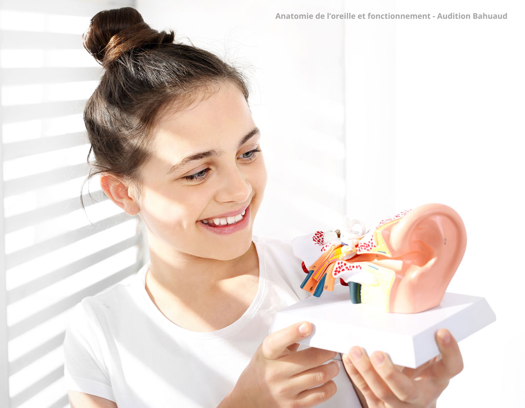
The outer ear is the visible part of the ear. It consists of two main structures:
- The auricle: made up of cartilage and covered with skin, the auricle acts like a funnel and is thus able to capture sound waves, identify the direction of sounds, amplify them and focus them so that they are directed towards the ear canal. Thanks to its shape, the pinna protects the middle and inner ear from particles and debris.
- The ear canal: measuring approximately 2.5 cm in adults, it is lined with skin, hairs and glands that produce earwax. It extends from the outer ear to the eardrum, thereby enabling the transmission and amplification of sound, protecting the eardrum, regulating temperature and humidity inside the ear, and preventing infections thanks to earwax.
The middle ear
In the anatomy of the ear, the ‘middle ear’ is situated between the outer ear and the inner ear. It consists of:
- The eardrum, or tympanic membrane: a thin membrane that receives sound waves from the ear canal and converts them into vibrations, which it transmits to the various ossicles of the middle ear. It is able to vibrate without allowing solids or liquids to pass through, thereby protecting the inner ear.
- The ossicles: three small bones that transmit vibrations received from the eardrum to the inner ear.From the one closest to the eardrum to the inner ear, they are: the malleus, the incus and the stapes.
- The Eustachian tube: this is a passage connecting the middle ear to the nasopharynx, which serves to equalise air pressure on either side of the eardrum, ensuring normal hearing and protecting the eardrum.
The inner ear
The inner ear, which is invisible to the naked eye, is situated behind the middle ear, deep within the skull. This part of the ear’s anatomy plays a crucial role in hearing; it consists of:
- The cochlea: made up of sensory hair cells, it converts sound vibrations into nerve signals.
- The auditory nerve, or cochlear nerve: transmits nerve signals received from the cochlea to the brain, where they are perceived as sounds.
- The vestibule: contains two sensory organs, the utricle and the saccule. The vestibule detects the position of the head in relation to gravity and plays a central role in balance.
- The semicircular canals: three canals containing sensory hair cells that detect rotational movements of the head to maintain balance.
- The vestibular nerve: transmits information about balance received from the vestibule and the semicircular canals to the brain.
Our hearing care professionals are committed to offering you solutions tailored to your individual needs and lifestyle – find out more about Audition Bahuaud’s commitments.
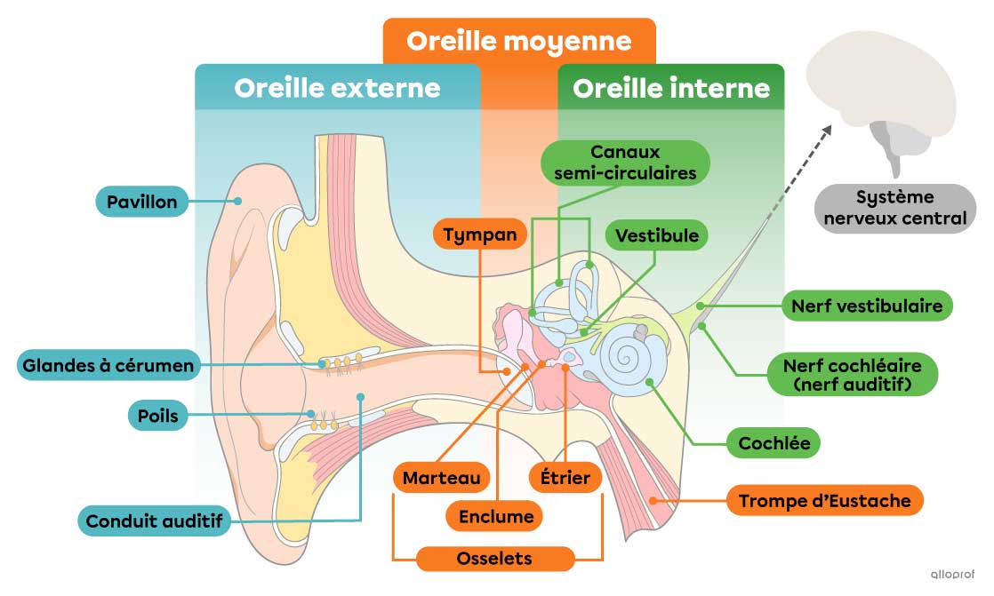
Diagram of the anatomy of the ear
Understanding the anatomy of the ear is essential for grasping the mechanisms of hearing and balance. In this diagram of the ear, we can see the three parts – the outer ear, the middle ear and the inner ear – as well as all their components, which we have described in detail earlier in the article
How does hearing work?
By studying the anatomy of the ear, we can understand how hearing works. To summarise the various elements we have looked at previously, hearing begins with the outer ear capturing sound waves. The middle ear then amplifies and transmits these sounds to the inner ear, which converts them into electrical signals sent to the brain. Finally, the brain interprets these sounds, such as human speech, music, environmental noises, etc. Discover our hearing centres near Toulouse and enjoy a free hearing test
What is the most delicate part of the ear?
The most fragile part of the ear is the cochlea. Located in the inner ear, the cells that make up the cochlea are highly sensitive to damage caused by infections, excessive noise, ageing, etc.As these cells do not regenerate once damaged, the damage is irreversible. Since the cochlea plays a crucial role in transmitting signals to the brain, damage to these cells can lead to total hearing loss.
Common ear problems
The anatomy of the ear is designed to provide the best possible protection for all its parts. With effective prevention and high-quality care, it is possible to reduce the risk of common conditions such as:
- Infections and inflammation: external otitis and internal otitis
- Hearing problems: hearing loss and tinnitus
- Barotrauma (injuries resulting from a rapid change in pressure)
- Earwax blockages
Find your hearing care professional to help with your hearing problems.

How can you protect your ears?
To protect your ears as effectively as possible, it is advisable to practise good hygiene by cleaning your ears regularly and avoiding cotton buds, as well as keeping your ears dry after swimming to prevent moisture-related infections.
When travelling by plane or going scuba diving, remember to equalise the pressure by chewing, yawning or using earplugs designed specifically for these activities.
To look after your hearing, use hearing protection (earplugs, noise-cancelling headphones, etc.), turn down the volume on noisy devices (music, television, etc.), and take breaks when noise levels are high. Above all, to protect your ears and your hearing, make sure you see hearing specialists – such as ENT doctors and hearing aid specialists – regularly.
To make sure you don’t miss our news and product offers, follow us on Facebook and Instagram or check out our news here